dataset information -  licence  -  download  -  citation


An Early Experience Toward Developing 
Computer Aided Diagnosis for Gram-Stained Smears Images. 

Gram stained direct smears test is clinically useful in early identification of infections. However, this practice is time consuming and labour intensive. Most existing effort in this area is to perform high-magnification analysis of images taken from manually selected areas. We address the problem of the automatic selection of areas based on low-magnification images, where bacteria are likely to be found when viewed in high-magnification. In order to stimulate the interest in the community on this problem, we propose a novel benchmark evaluation dataset. The dataset comprises 1,600 images which were extracted from various body parts of patients with different medical conditions.
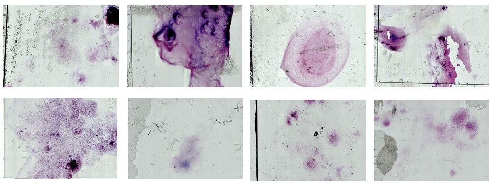

Dataset containing 8 Gram-stain slide images. Images are captured using a 2.5x objective (low-magnification).


Dataset Information

The specimen slides for the dataset were prepared at the Sullivan Nicolaides Pathology laboratory, Australia. The slides were then scanned at the University of Queensland. All the patient data had been de-identified prior releasing the dataset.

-Current Description (June 2017)
  • 8 Gram stain slide images.
  • Hardware: Zeiss motorised microscope & PixeLINK camera.
  • 200 patches randomly selected per image.
  • A total of 1, 600 patches.
  • 3 subjects labelled each patch. Based on that observation, each subject assigned a label:
    • four: best candidate area
    • three: pathologists may look if no bacteria are seen in areas with labels four
    • two: dense and dark area, and
    • one: background or unwanted artefacts (dirt, dust, oil).
  • All subjects agreed for 1,293 images. We decided to discard the remaining 307 images in order to create an unbiased dataset. These images often contained out-of-focus or incorrectly stained areas where the subjects had different opinions on their usefulness.


-Evaluation Protocol

We use the leave-one-out cross validation approach (LOOCV). The LOOCV protocol takes out all the patches from one Gram stain slide image for testing and uses all patches from the seven remaining Gram stain slide images for training. This is executed for every testing image in a rotating basis, and the overall accuracy is obtained by averaging the accuracy of all validations.


Licence

The Gram Stain Dataset and associated data ('Licensed Material') are made available to the scientific community for non-commercial research purposes such as academic research, teaching, scientific publications or personal experimentation. Permission is granted to you (the 'Licensee') to use, copy and distribute the Licensed Material in accordance with the following terms and conditions:

  • Licensee must include a reference to the following publication in any published work that makes use of the Licensed Material:

An Early Experience Toward Developing Computer Aided Diagnosis for Gram-Stained Smears Images
J. Carvajal, D. F. Smithy, K. Zhao, A. Wiliem, P. Finucane, P. Hobson, A. Jennings, R. McDougall, and B. Lovell
Computer Vision and Pattern Recognition (CVPR) Workshop on Computer Vision for Microscopy Image Analysis (CVMI), 2017.

  • If the Licensee alters the content of the Licensed Material or creates any derivative work, Licensee must include in the altered Licensed Material or derivative work prominent notices to ensure that any recipients know that they are not receiving the original Licensed Material
  • Licensee may not use or distribute the Licensed Material or any derivative work for commercial purposes including but not limited to, licensing or selling the Licensed Material or using the Licensed Material for commercial gain
  • The Licensed Material is provided 'AS IS', without any expressed or implied warranties. The authors do not accept any responsibility for errors or omissions in the Licensed Material
  • This original licence notice must be retained in all copies or derivatives of the Licensed Material
  • All rights not expressly granted to the Licensee are reserved by the authors

Download

The current version of the Gram Stain Dataset [download].


Acknowledgements

This work has been funded by Sullivan Nicolaides Pathology, Australia and the Australian Research Council (ARC) Linkage Projects Grant LP130100230.


Citation

 J. Carvajal, D. F. Smith, K. Zhao, A. Wiliem et al. "An Early Experience Toward Developing Computer Aided Diagnosis for Gram-Stained Smears Images".  Computer Vision and Pattern Recognition (CVPR) Workshop on Computer Vision for Microscopy Image Analysis (CVMI), 2017. [pdf]